 Anatomy and Screening Programmes
It is estimated that the NHS Cervical Screening Programme saves the lives of 5000 women in the UK each year
1964 saw the introduction of an initially primative screening service, providing cervical cell analysis but failing to target those most at risk. Despite this, the introduction of the NHS Cervical Screening Programme (NHSCSP) in 1998 was quick to evolve and rapidly introduced the call-recall system, by which women are automatically invited to attend for screening. Today, this stands at every 3-5 years from the age of 25 until 65 with an attendance rate just under the governmental target of 80%.
The Screening Process
The Papanicolaou Test, or "Pap Smear" is predominantly used as a preventative measure, rather than a cure for cervical cancer. The Liquid Based Cytology (LBC) method is able to indicate abnormal cellular changes to the cervical tissue before the transformation into a malignant tumours, the most common forms being squamous cell carcinoma (2/3) and adenocarcinoma. Thus the regularity of screening is necessary, as the first stages of cervical cancer are often asymptomatic.
The cervix, part of the female reproductive system, is classed as the conical collection of cells forming the narrowing, or "neck" of the uterus before joining the vagina. A sample of cells is collected from the external os (orifice) and an area known as the Transformation Zone. The latter region represents the border between the simple, columnar cells of the inner endocervix and stratified squamous cells of the outer ectocervix, and it is the highly metaplastic nature of these cells which can result in pre malignant or malignant changes.
These cells are prepared for visualisation by light microscopy via the LBC method, in which the distal end of the cell collection device is stored in a liquid preservative and spun at high speed within a laboratory to remove pollutants, such as blood cells from the sample. Prior to the introduction of this method in 2008, 9% of samples were deemed inadequate due to poor cell visibility, however this has subsequently fallen to just 2.5%, dramatically reducing patient anxiety.
Results
In 9/10 cases, the cervical cells are normal, but in 1/10 samples, the cells show dysplasia, often characterised according to the Cervical Intraepithelial Neoplasia (CIN) scale, depending on the severity of the premalignant transformation or abnormal growth. Further investigations and treatment are required for both CIN 2 and 3 stages, for which more information can be found on the Cancer Research UK website.
Note, that CIN 3, otherwise known as "carcinoma in situ" is a small, localised malignancy that has yet to affect the basement membrane of the tissue, so is technically classed as a non-invasive cancer.
That does not mean however, that cases of invasive cervical cancer are not detected through the NHSCSP. This is a very rare occurrance, affecting just 2/10,000 and of those, 56% go on to be diagnosed with cervical cancer.
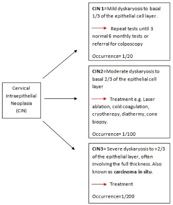
Image of Anatomy of Uterus and Cervix courtesy of Wikimedia Commons. Public domain.